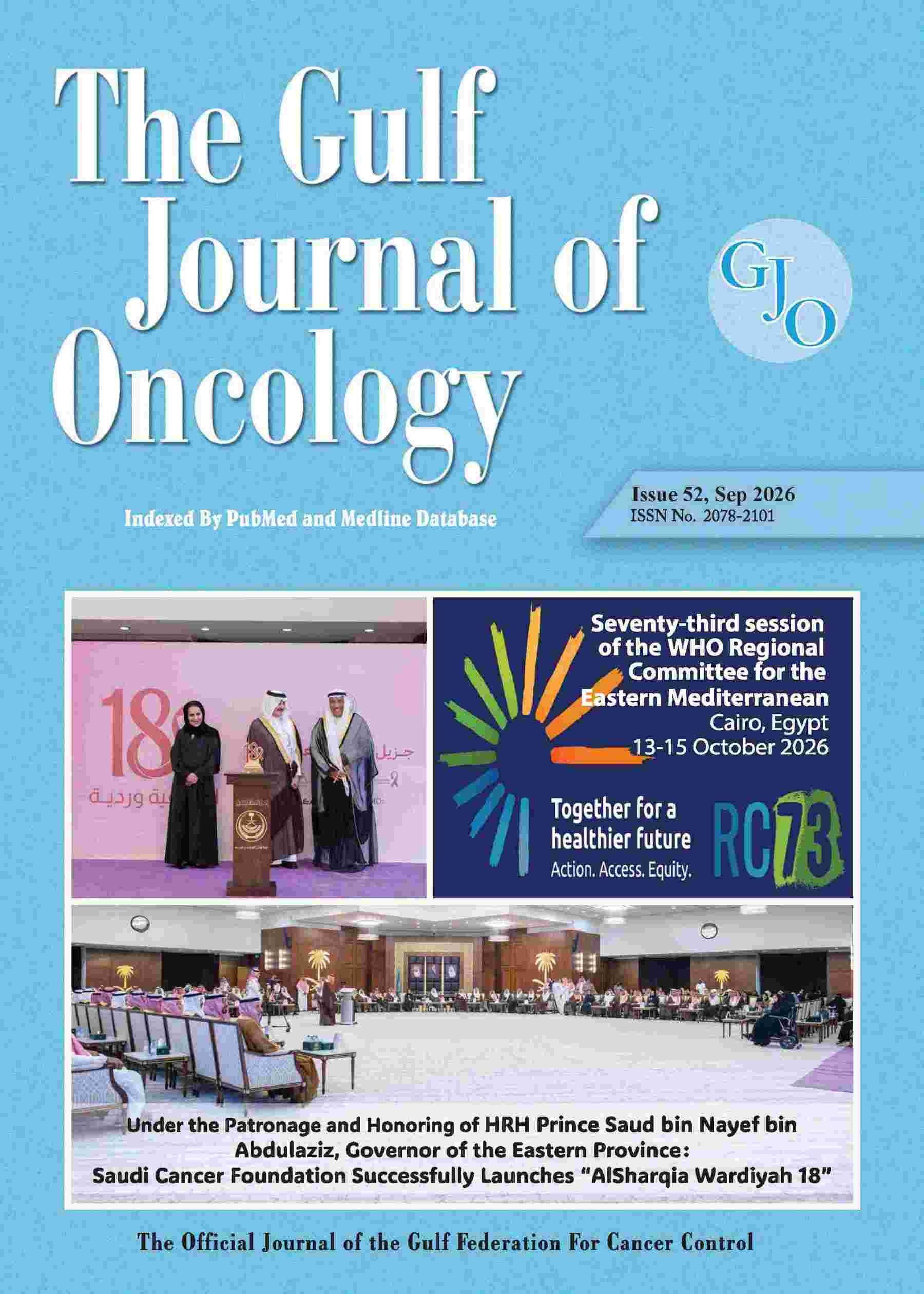
 Issue No. 52, Sep 2026 Electronic ISSN 2521-3881 |
||
| Issue No. 52 - Sep2026 | ||
| 7-13 | Clinico-Demographic Variables Affecting Survival in Esophageal Cancer: Real-World Evidence from a Tertiary Cancer Centre in Western India | |
|
Samiran Chavan, Atul Kumar Gupta, Bharti Devnani, Puneet Pareek Department of Radiation Oncology, All India Institute of Medical Sciences, Jodhpur, Rajasthan, India |
||
| Abstract | ||
|
Introduction or Background: Esophageal cancer (EC) is a major global health concern, ranking seventh in incidence and sixth in cancer-related mortality. In India, it accounts for approximately 47,000 new cases and 42,000 deaths annually. Squamous cell carcinoma (ESCC) is the predominant histology, particularly among socioeconomically disadvantaged populations. Most patients present with advanced disease and poor performance status, limiting curative treatment options. While trials such as CROSS and RTOG 85-01 have established the benefits of multimodal therapy, real-world outcomes from low-resource settings like Western Rajasthan remain underrepresented. This study aimed to evaluate overall survival and identify prognostic factors in patients with EC treated at a tertiary center in rural India.
Materials and Methods: A retrospective observational study was conducted at the Department of Radiation Oncology, AIIMS Jodhpur, analyzing data from January 2020 to December 2023. Patients with histologically confirmed ESCC or adenocarcinoma who received definitive treatment were included. Data on demographics, ECOG performance status, tumor site, clinical stage, treatment modality, and survival outcomes were extracted. Overall survival (OS) was calculated from the date of diagnosis to death or last follow-up. Kaplan-Meier curves were used for survival analysis, and the log-rank test evaluated subgroup differences. Variables significant in univariate analysis were subjected to multivariate Cox regression.
Results: Out of 61 patients, the median age was 58 years, with 32 females and 29 males. Surgical resection was performed in 24 patients; 40 received concurrent chemoradiotherapy. Median OS was significantly higher in the surgical group (95 vs. 13 months, p=0.001) and in those receiving concurrent chemotherapy (24 vs. 12 months, p=0.021). Performance status was a strong predictor: ECOG 0 and 1 had superior OS compared to ECOG 2 and 3 (p=0.004). Local recurrence was associated with poorer outcomes (p<0.001), while distant metastasis did not significantly impact OS (p=0.639). Clinical stage showed a trend toward significance (p=0.064). On multivariate analysis, surgery (HR 0.30, p=0.001), concurrent chemotherapy (HR 0.46, p=0.019), and absence of local progression (HR 0.31, p=0.020) were independent predictors of improved survival.
Discussion: The study highlights the survival benefit of multimodal treatment, particularly surgery and concurrent chemoradiotherapy, in esophageal cancer. Patients with good performance status were more likely to receive and tolerate intensive treatment, resulting in better outcomes. The findings align with global evidence supporting trimodality therapy for resectable, locally advanced disease. Poor outcomes in patients with ECOG =2 or local progression emphasize the need for early detection, nutritional support, and prehabilitation to optimize fitness for curative therapy.
Conclusion: In this real-world study from a rural Indian cancer center, surgery and concurrent chemoradiotherapy significantly improved survival in esophageal cancer. Performance status and local disease control emerged as key prognostic factors. These findings underscore the need for timely diagnosis, patient optimization, and access to multidisciplinary care to improve outcomes in resource-constrained settings. Prospective, multicenter studies are warranted to further refine treatment strategies tailored to low- and middle-income countries.
Keywords: Esophageal cancer, concurrent chemoradiotherapy, overall survival, prognostic factors
|
||
| full article | ||
| 2 | 14-19 |
Characteristics of hospital readmissions within 30 days among adult patients with cancer in Jordan |
|
Nour M. Mustafa1, Pharm D, BCOP, Dima Attili1, Pharm D, Nour Faqeer1, Pharm D, BCOP 1 Department of Pharmacy, King Hussein Cancer Center, Amman, Jordan |
||
| Abstract | ||
|
Background: Hospital readmissions are common among cancer patients, related to the complexity of their disease. This study aims to describe the characteristics of unplanned hospital readmission in cancer patients.
Methods: This prospective study was conducted at King Hussein Cancer Center from March 2023 to August 2023. The study included unplanned hospital readmission, defined as any admission through the emergency room within 30 days following the initial admission, excluding patients admitted to palliative care. Patients were followed to collect baseline characteristics, and data related to readmission.
Results: During the study period, a total of 2435 unplanned admissions were included. Of these, 769 (31.6%) patients had at least one readmission, accounting for a total number of 963 readmissions. The mean age of the readmitted patients was 55.9 years (± 14.6 SD), with 49.9% being male. The most common cancer types among these patients were gastrointestinal (22.4%), hematological (21.2%), and breast cancer (16.5%). Additionally, 80% had metastatic disease, and 54.4% were receiving active antineoplastic treatment. Infections were the leading cause of initial admission (28.3%) and first readmission (31.6%). Notably, 36.2% of the patients were readmitted for the same reason as their initial admission. The median time to first readmission was 11 days (range: 1-44 days). Among readmissions, 6.8% required intensive care unit service, while 89.2% were directed to the medical oncology service.
Conclusion: Unplanned readmissions within 30 days among patients with cancer were common, with infection being the leading cause. Further studies are needed to reduce preventable readmissions to optimize patient care.
Key words: Hospital admission, Hospital readmission, cancer, discharge.
|
||
| full article | ||
| 3 | 20-28 | Original Article | Efficacy of Volumetric Modulated Arc Therapy (VMAT) in Nasopharyngeal Carcinoma: A Retrospective Analysis |
|
Venkada Manickam Gurusamy1, Palmira Caparrotti1, Hissa Hussain Al-Abdulla1, Saju Raveendran Divakar1, Ghazia Shaik1, Patsey Conway-Murray1, Amna Rafi Vora1, Muhammad Usman1, Rabih Wafiq Hammoud1, Noora Al-Hammadi1 1 Department of Radiation Oncology, National Center for Cancer Care and Research (NCCCR), Hamad Medical Corporation, Doha, Qatar. |
||
| Abstract | ||
|
Purpose: This retrospective study evaluates the efficacy of volumetric arc radiotherapy (VMAT) in NPC patients at a tertiary hospital in Qatar, serving a diverse population. The primary endpoint was loco-regional tumor control, while secondary endpoints included disease-free survival, overall survival and treatment-related toxicities.
Methods and materials: Patients older than 16 years who had T1 - T4N0 - 3M0 nasopharyngeal cancer and the WHO performance status of 0 or 1 were included. All patients received radiotherapy with or without chemotherapy as indicated. Initially, our institution policy was concurrent chemoradiotherapy (CCRT) followed by adjuvant chemotherapy for advanced stage disease. After the publication of randomized trials showing significant improvement in clinical results with induction chemotherapy followed by concurrent chemoradiation, our practice was changed accordingly.
Results: All 67 patients completed volumetric arc intensity-modulated radiotherapy and the overall median radiotherapy dose was 70 Gy (IQR 66 - 76 Gy). In total, 12 of 67 patients (17.9%) received CCRT alone and 52 of 67 patients (77.6%) received either induction chemotherapy or adjuvant chemotherapy. We recorded a total of 11 events of recurrence, distant metastasis or death in 8 of 67 patients (11.9%). Distant metastasis was seen in 6 of 67 patients (89.5%), and 3 of 67 patients (4.6%) had both locoregional failure and distant metastasis. For all patients, 3-year overall survival (OS), disease-free survival (DFS) and locoregional recurrence-free survival (LRRFS) were 98% (95% CI, 94.1 - 100%), 87.3% (95% CI, 78.6 - 97%) and 92.8% (95% CI, 86.1 - 99.9%), respectively.
Conclusion: Our analysis demonstrates that the combination of VMAT and chemotherapy achieves excellent long-term tumor control, with outcomes comparable to previously published data.
|
||
| full article | ||
| 4 | 29-43 |
Prevalence, Correlates, and Predictors of Depression and Anxiety Among Cancer Patients in the UAE: A Comprehensive Cross-Sectional Study Using the Hospital Anxiety and Depression Scale (HADS) |
|
Manal Ali Buabeid1, Ayesha Alqasem1, Maitha Salem Althehli1, Shamma Ali Alhefeiti1, Faryal Iqbal2, Amin Abyad2, Hassan Jaafar2, Humaid O. Al-Shamsi2-9* 1 Fatima College of Health Sciences, Department of Pharmacy, Abu Dhabi, United Arab Emirates 2 Burjeel Cancer Institute, Burjeel Medical City, Abu Dhabi, United Arab Emirates 3 Nexomic, Dublin, Ireland 4 Department of Medical Oncology, Dana-Farber Cancer Institute, Harvard Medical School, Boston, MA, United States 5 Harvard Medical School, Harvard University, Boston, MA 20138, United States 6 College of Medicine, Ras Al Khaimah Medical and Health Sciences University, Ras Al Khaimah, United Arab Emirates 7 Gulf Medical University, Ajman, United Arab Emirates 8 Emirates Oncology Society, Dubai, United Arab Emirates 9 College of Medicine, University of Sharjah, Sharjah, United Arab Emirates |
||
| Abstract | ||
|
Background: Depression and anxiety are highly prevalent among cancer patients, yet comprehensive data on their prevalence, correlates, and predictors in the Middle East remain limited. Understanding these factors is crucial for developing effective psycho-oncological interventions in culturally diverse healthcare settings. Objective: To determine the prevalence of depression and anxiety among cancer patients in the UAE, to examine correlates of psychological distress, and to identify predictors of mental health service utilization using the Hospital Anxiety and Depression Scale (HADS). Methods: A comprehensive cross-sectional analysis was conducted among 105 cancer patients at a major UAE healthcare facility (Burjeel Cancer Institute, Burjeel Medical City) between April and August 2025. Participants completed structured questionnaires in English or Arabic, including demographic information, cancer-related factors, and the HADS. Pearson correlation analyses examined relationships between variables and psychological distress. The logistic regression analyses identified predictors of psychological care and medication utilization. Results: The study revealed high prevalence rates of psychological distress: 60.0% of participants scored in the clinical range for depression (HADS-D ≥11) and 43.8% scored in the clinical range for anxiety (HADS-A ≥11). A strong correlation was observed between anxiety and depression scores (r = 0.742, p < 0.001). Treatment side effects showed consistent moderate correlations with both anxiety (r = 0.284, p = 0.003) and depression (r = 0.319, p = 0.001). Anxiety demonstrated stronger correlations with service utilization than depression (psychological care: r = 0.356 vs. r = 0.298; medication use: r = 0.421 vs. r = 0.389). In multivariate regression analysis, treatment side effects significantly predicted psychological care utilization (OR = 3.45, 95% CI: 1.12-10.62, p = 0.031), while psychological care receipt strongly predicted medication use (OR = 8.45, 95% CI: 2.89-24.73, p < 0.001). Despite high distress levels, only 21.9% received psychological care and 18.1% used psychiatric medications, while 65.7% expressed interest in additional mental health support. Conclusion: This comprehensive analysis reveals substantial psychological distress among UAE cancer patients, with depression affecting three in five patients and anxiety affecting two in five. Strong anxiety-depression comorbidity supports integrated screening approaches. Treatment side effects emerged as key predictors of service utilization, highlighting the interconnection between physical and psychological symptoms. Significant treatment gaps exist, with most patients expressing unmet mental health needs. Healthcare systems should prioritize routine HADS screening, systematic side-effect assessment, and culturally sensitive, integrated psycho-oncological services to address this substantial burden of psychological distress. Keywords: cancer; depression; anxiety; HADS; correlation analysis; regression analysis; psycho-oncology; UAE; mental health service utilization; treatment predictors |
||
| full article | ||
| 5 | 44-51 | Induction spatially fractionated GRID radiotherapy (SFGRT) radiotherapy for locally advanced extremity soft tissue sarcoma: Report of 10 years follow up. |
|
Abdullah Alsuhaibani1, Mohammed Almahmoud2, Mohammed Mohiuddin2, Muhammad Memon3, Ahmed Nobah4, Medhat Elsebaie2,6, Ayman Elshenenawy3,5, Rajive Pant4, Mahmoud Shaheen4, Majid Alyamani4, Hatim Khoja4, Tariq Alzaid4, Fouad Al Dayal4. Ahmed Elashwah2,5 1 King Khaled university hospital, King Saud University, oncology center, Riyadh Saudi Arabia. 2 King Faisal specialized hospital and research center, oncology center, radiation oncology section, Riyadh, Saudi Arabia. 3 King Faisal specialized hospital and research center, oncology center, medical oncology section, Riyadh, Saudi Arabia. 4 King Faisal specialized hospital and research center, Riyadh, Saudi Arabia. 5 Kasr Al-Eini Center of Clinical Oncology (NEMROCK), Cairo University, Cairo, Egypt. 6 Department of radiation oncology, National Cancer Institute (NCI), Cairo University, Cairo, Egypt. |
||
| Abstract | ||
|
Introduction: Increasing the rate of tumor necrosis by more than 90% for extremity soft tissue sarcoma (STS) with preoperative treatment has been associated with better local control and survival. Locally advanced tumors appears to be more challenging to achieve the desired necrosis rate (>90%) with preoperative chemoradiation. We aim to test a novel approach using induction spatially fractionated GRID radiotherapy (SFGRT) followed by preoperative chemoradiation in the treatment of STS looking for improved tumor necrosis rate and to report survival data after 10 years follow up. Patients and Methods: Fourteen patients with locally advanced bulky extremity STS were included in the study. Median tumor size is 11.5 cm (8-26 cm). All patients were treated with induction 18 Gy single fraction SFGRT followed by external beam radiotherapy 50 Gy / 25 fractions / 5 weeks concurrently with chemotherapy (Iphosphamide 2 gm/m2/day x 3 days and mesna 2 gm/day x 3 days in divided doses every three weeks for total of 2 cycles). Patients were planned for surgical resection 4-6 weeks after completing neoadjuvant treatment. Results: Twelve (86%) patients completed the planned treatment. One (7.1%) patient had treatment interruption due to grade 3 skin toxicity and 1 patient underwent an amputation of the foot mid of the neoadjuvant treatment. Thirteen (93%) patients underwent limb salvage surgical resection, one of them had positive surgical margins. Nine (65%) patients had > 90 % tumor necrosis with 2 patients having a pathological complete response. At a median follow up of 37 months, 3 patients (23%) lost their follow up with no evidence of local recurrence or distant metastasis. At 5 years follow up (median follow up 60 months), 3 (23%) patients had distant metastasis and died. At 10 years follow up (median follow up 92 months), 7 (53.8%) patients are alive with no evidence of local recurrence or distant metastasis. Conclusion: The addition of induction spatially fractionated GRID radiotherapy SFGRT to preoperative chemoradiation showed to be an effective way to improve the rate of tumor necrosis and enhancing response to chemoradiation resulting in improvement in local control and overall survival in our small cohort over a long follow up period. Using SFGRT and neoadjuvant CCRT appeared to be safe and should be tested on a larger population with locally advanced extremity STS. Key words: induction, radiotherapy, SGFRT, soft tissue sarcoma . |
||
| full article | ||
| 6 | 52-64 | Modeling Cancer Incidence and Mortality in Yemen Using GLOBOCAN 2020 Estimates: An Empirical and Rank-Based Analysis |
|
Gamal Abdul Hamid1,2, Amen Bawazir3,4, Safa A. Shukry2, Taher Bin Sumiat5, Noureddine Ouerfelli6 1 Hematology and Laboratory Department. Faculty of Medicine and Health Sciences, University of Aden, Aden, Yemen 2 National Oncology Center, Aden, Yemen. 3 College of Medicine. AlMaarefa University. Riyadh, Saudi Arabia. 4 Community Medicine Department. Faculty of Medicine and Health Sciences, University of Aden, Aden, Yemen 5 Seiyun University. College of Medicine. Hadhramout. Yemen 6 Institute Supérieur des Technologies Médicales de Tunis, LR13SE07, Laboratoire de Biophysique et Technologies Médicales, Université de Tunis El Manar, Tunis, Tunisia. |
||
| Abstract | ||
|
Background: Cancer is a growing public health concern in Yemen, yet comprehensive quantitative analyses of its epidemiology remain limited. This study provides an empirical, rank-based model of cancer incidence and mortality in Yemen using GLOBOCAN 2020 estimates, aiming to characterize the relationship between new cases and deaths and to evaluate the applicability of linear, logarithmic, and power-law models. Methods: We analyzed GLOBOCAN 2020 data for Yemen, including 16,476 new cancer cases and 12,103 cancer-related deaths. Linear regression (forced through origin), logarithmic transformation, and power-law models were applied to model the relationship between the number of new cases (Nc) and deaths (Nd). A severity index (s) derived from rank order was introduced, and cumulative risks were modeled. Comparisons with global and regional data were performed. Results: Breast cancer was the most frequent malignancy (17.6% of cases), followed by colorectal cancer, leukaemia, stomach cancer, and non-Hodgkin lymphoma. The linear model yielded a mortality-to-incidence ratio of 67.74% (Nd = 0.6774 x Nc). The logarithmic model provided an improved fit (lnNd = 1.04565 x lnNc - 0.62732; R ≈ 0.986), indicating a near-unity behavioral index (a = 1.04565). A power-law model (Nd = 0.53402 x Nc1.04565) further confirmed this robust dependency. High-lethality cancers (oesophagus, liver, pancreas, and lung) showed mortality rates approaching 95%, whereas breast and thyroid cancers had lower ratios (56.6% and 26.3%, respectively). Rank-based analyses revealed exponential decay patterns, and a novel severity metric (s) effectively stratified cancer burden. Conclusions: The strong correlation between incidence and mortality in Yemen underscores a high overall cancer lethality, likely due to late-stage diagnosis and limited treatment access. Mathematical modeling, including power-law and severity indices, provides actionable insights for resource allocation and early detection priorities. Strengthening cancer registries and expanding screening programs are critical. Keywords: GLOBOCAN Yemen 2020, cancer epidemiology, incidence and mortality, empirical modeling, rank-based analysis, severity metric, cumulative risk, age-standardized rates, power-law model. |
||
| full article | ||
| 7 | 65-78 | Establishing the First Four-Dimensional Multipurpose Central Nervous System Tumors Biobank in Saudi Arabia: Project Design, Perspectives, and Preliminary Results |
|
Deema Hussein1,2, Saleh Baeesa3,4*, Alazouf Alhowity1, Ishaq Khan1,5, Mona Alomari1, Hanadi Qashqari1, Nawal Madkhali1, Reem Alserihi1,6, Mohammed Bangash4, Fahad Alghamdi7, Awatif Jamal7, Mohamad Saka1,2, Torki AlZughaibi1,2, Ghazi Damanhouri1, Adeel Chaudhary8, Adel Abuzenadah1,2,8 1 King Fahd Medical Research Center, King Abdulaziz University, Jeddah 21589, Saudi Arabia. 2 Department of Medical Laboratory Sciences, Faculty of Applied Medical Sciences, King Abdulaziz University, Jeddah 21589, Saudi Arabia. 3 Neurosciences Department, King Faisal Specialist Hospital and Research Center, Jeddah, Saudi Arabia. 4 Division of Neurosurgery, Faculty of Medicine, King Abdulaziz University, Jeddah 21589, Saudi Arabia. 5 MIT Media Laboratory, Massachusetts Institute of Technology, 75 Amherst St., Cambridge, MA 02139, United States. 6 College of Health Sciences, Al-Rayan Colleges, 41411 Madinah Al-Munawarah, Saudi Arabia. 7 Pathology Department, Faculty of Medicine, King Abdulaziz University, Jeddah 21589, Saudi Arabia. 8 Center of Innovation for Personalized Medicine, Department of Medical Laboratory Technology, Faculty of Applied Medical Sciences, King Abdulaziz University, Jeddah 21589, Saudi Arabia. |
||
| Abstract | ||
|
Introduction: The burden of central nervous system tumors (CNSTs) is a global challenge. Disease-associated biobanks enable the use of collected human tumor cell samples to improve the understanding of disease biology and advance personalized medicine. However, differences in genetic composition can influence drug treatment outcomes; thus, variable samples collected from different genetic populations are necessary. Currently, tumor cell line data derived from CNSTs related to the Middle Eastern genetic pool are rare in the literature. Therefore, we established a CNSTs biobank that focuses on initiating CNSTs research in vitro in Saudi Arabia. Materials and Methods: A total of 111 fresh CNSTs specimens were collected between February 2013 and December 2020 from patients who were treated at King Abdulaziz University Hospital in Jeddah, Saudi Arabia. The collection procedure, tissue processing, and in vitro primary cell culture initiation procedures are detailed. Results: The characteristics of patients and samples collected in the biobank showed a female-to-male ratio of 61:39%, a mean age of 39.7 years, and variable tumor types, including low-grade, high-grade malignant, and metastatic brain tumors. The basic characteristics of tumor tissue samples, including tumor volume, composition, hydration, and vascularization, were not defined by their origin. Primary tumor cell lines were established for histopathologically distinct tumor types, and the observed cells exhibited variable morphologies. Conclusion: We present the first four-dimensional multipurpose CNSTs biobank protocol design in Saudi Arabia, along with our preliminary results. The cohort study encompassed a diverse range of histologically variable CNSTs, including adult and pediatric cases. By establishing this biobank, we intend to foster national and regional collaborations to enhance translational research and targeted medicine for patients with CNSTs. We also encourage researchers to establish similar biobanks in their respective countries. Keywords: Cancer, biobanks, central nervous system tumors, primary cell lines, Saudi Arabia. |
||
| full article | ||
| 8 | 79-91 | Review Article | Craniospinal Irradiation (CSI) Techniques: A Systematic Review of Comparative Dosimetric Outcomes, Organ-at-Risk Sparing, and Treatment Efficiency |
|
Kesava Ramgopal Adavikolanu1, Chandramouli Ramalingam2, Soorej Balan Kaliyath1, Abhishek Raghava KS3, Thirumal M2, Nagasai Divya Kari2, Pragnahitha Neerudu2, Ambedkar Yadala4, Ajay Kumar Kondeti2 1 Department of Radiation Oncology, All India Institute of Medical Sciences (AIIMS), Mangalagiri, India |
||
| Abstract | ||
|
Background: Craniospinal irradiation (CSI) is technically demanding because the treatment volume extends along the entire neuraxis. Adequate coverage has to be maintained over a long target while limiting junction-related uncertainties and unnecessary irradiation of normal tissues. Although several techniques are now used for CSI, their relative dosimetric strengths and limitations are not fully established. Methods: We performed a systematic review based on a registered PROSPERO protocol. PubMed/MEDLINE, Embase, and Scopus were searched for comparative, patient-based CSI studies published between January 2000 and February 2026. The review focused on target coverage, conformity index (CI), homogeneity index (HI), junction characteristics, organ-at-risk (OAR) doses, integral dose and low-dose exposure, monitor units (MU), and beam-on time (BoT). Risk of bias was assessed using domains adapted for comparative dosimetric studies. Results: Twenty-two (22) comparative studies were included. Contemporary IMRT, VMAT, and helical techniques generally achieved at least 95% target coverage. In a multicentre comparison, PTV V95 ranged from 97.8% to 99.8%, while CI improved from 0.6 with 3DCRT to 0.9 with VMAT. Proton therapy produced substantially lower low-dose exposure; esophageal V20 was 3.89% versus 65.87% and heart V20 was 0.03% versus 2.80% compared with photon therapy. In the comparison of TrueBeam 6-MV FFF, Halcyon 6-MV FFF, and IMPT, CTV D95 was 2367, 2375, and 2360 cGy, respectively, while total normal-tissue V1 Gy was 57.3%, 55.8%, and 17.8%. Mean heart dose was 291, 378, and 27 cGy, and mean lung dose was 505, 481, and 160 cGy, respectively. Halcyon reduced beam-on time to 6.1 minutes compared with 12.1 minutes for TrueBeam, while IMPT required 4.2 minutes. Conclusion: Modern photon techniques can achieve reliable CSI coverage while making junction management and treatment delivery more practical. Proton therapy offers a clear advantage in reducing low-dose and integral-dose exposure. In practice, the choice of technique should take into account the patient's anatomy and age, the treatment goal, available equipment, and the resources and expertise of the treating centre. Key Words: Craniospinal irradiation; Dosimetric comparison; Photon radiotherapy; Proton therapy; Organ-at-risk sparing |
||
| full article | ||
| 9 | 92-97 | Tubular Ileal Duplication Associated with Diffuse Polyposis and Metastatic Adenocarcinoma Presenting as Acute Small Bowel Obstruction in an Adult: First Reported Case and Review of the Literature |
|
Aliasgar I Kachwala1, Obaid M Alharbi1, Rajan Arora2 1 Department of general surgery, Farwaniya hospital, Kuwait |
||
| Abstract | ||
|
Tubular duplication of the alimentary tract is rare during adulthood. Most of duplications are cystic in nature. Malignant changes in cystic duplication had been described in literature. We encountered male in his 50,s with no previous significant complaints presented with acute intestinal obstruction. On diagnostic laparoscopy found to have multiple peritoneal, omental deposits with transition zone at narrowed junction of duplicated loop with ileal loop. Resection and primary anastomosis with uneventful postoperative recovery was achieved. Postoperative histopathology examination revealed metastatic adenocarcinoma stage pT4N2M1. Interestingly tubular duplicated segment was blind at proximal end and contained multiple mucosal polyps (more than 100 in number) which were small, sessile and mostly range in size from 0.1 to 0.4 cm in diameter while the largest polyp measured 0.7 cm and was located immediately proximal to tumor. Based on available research we hypothesize that pathogenesis in our patient was driven by the unique anatomy and microenvironment of the tubular duplication segment. A milieu of blind proximal end of the duplicated loop combined with heterotropic gastric mucosa, delayed transit times, accumulation of abnormal non intestinal secretions and consequent bacterial overgrowth lead to state of chronic persistent mucosal inflammation. This combined insult resulted in mucosal hyperplasia forming polyps and subsequent dysplastic changes that ultimately transitioned into adenocarcinoma suggesting adenoma carcinoma sequence. Whenever encountered, complete surgical resection of the duplicated segment should be undertaken due to high probability of dysplasia and malignancy transformation of mucosal lesions as highlighted in this case report. Keywords: Tubular ileal duplication, Intestinal polyposis and malignant transformation, intestinal obstruction, Adenocarcinoma, Adult. |
||
| full article | ||
| 10 | 98-102 | Case Report | Tubulolobular Carcinoma of the Breast: First Case Report from Saudi Arabia |
|
Mehad Ahmed1, Wafaey Badawy2, Samah O. Mohager1 1 Prince Sattam Bin Abdulaziz University, Department of Basic Medical Sciences, College of Medicine. AlKharj, Riyadh, Saudi Arabia. |
||
| Abstract | ||
|
Tubulolobular carcinoma (TLC) of the breast is a singular invasive carcinoma with dual histologic features of both tubular and lobular elements within the same lesion. We report a right breast lump for 2 month in a 68-year-old lady. A simple mastectomy of the right breast was done for her and the surgical pathology report confirmed the diagnosis of tubulolobular carcinoma. This is the first documentation of the clinicopathologic features of this tumor in Saudi Arabia. Examination of the literature shows that to date, agreement on the classification of this tumor has not been reached, and although immunohistochemistry has a role, the histomorphologic features are still the mainstay for diagnosis. Keywords: breast tumor, case report, Tubulolobular carcinoma, histopathology . |
||
| full article | ||